


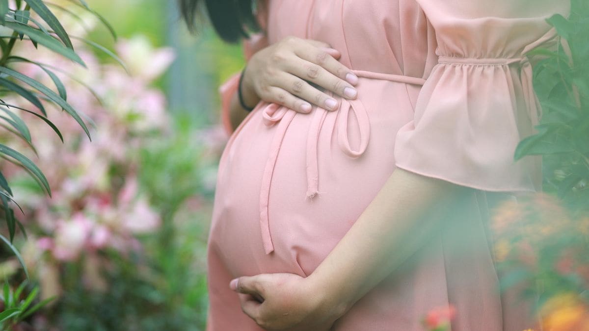
Doctors in Bulandshahr, Uttar Pradesh, were left stunned after an MRI scan of a 30-year-old woman revealed something highly unusual, she was 12 weeks pregnant, but the foetus wasn’t in her uterus. It was growing in her liver.
The woman had unknowingly developed intrahepatic ectopic pregnancy, a condition so rare that many doctors go their entire careers without encountering it. Experts believe this could be the first such case ever documented in India.
Here’s what we know so far about this baffling medical case, how it was discovered, and what it means for the patient moving forward.
What is intrahepatic ectopic pregnancy?
An ectopic pregnancy occurs when a fertilised egg implants itself outside the uterus, most commonly in the fallopian tubes. But in this case, the foetus made its way to the liver, a scenario that medical experts say is extremely rare.
When a pregnancy develops inside the liver, it’s termed an intrahepatic ectopic pregnancy, a condition so uncommon that only eight such cases have ever been reported worldwide so far, in countries including China, Nigeria, the United States, and parts of Europe.
Dr KK Gupta, a radiologist at a private imaging centre in Meerut, who uncovered it while carrying out the MRI abdomen test of the woman, told News18, “When I saw the scan, I could not believe my eyes. The foetus was embedded in the right lobe of the liver, and there were clear cardiac pulsations. I have never seen such a case in my career, and according to available data, this might be India’s first intrahepatic ectopic pregnancy.”
How did the case come to light?
The breakthrough came after the woman endured weeks of persistent abdominal pain and vomiting. With no clear answers from initial tests, doctors decided to go a step further and advised an MRI scan of the abdomen, a test typically used when ultrasound results are inconclusive.
The scan was carried out at a private imaging centre in Meerut, under the supervision of Dr KK Gupta, a senior radiologist known for his decades of experience in advanced imaging.
Unlike a regular ultrasound, the MRI offered detailed, high-resolution images of the abdominal organs, layer by layer. What it uncovered left even seasoned professionals stunned.
“We observed a well-formed gestational sac inside the right lobe of the liver,” Dr Gupta told News18. “The foetus measured approximately 12 weeks in gestational age. Most strikingly, the scan confirmed active cardiac pulsations, establishing that the foetus was alive. At the same time, the uterus was completely empty, ruling out a normal intrauterine pregnancy,” he explained.
Dr Gupta further detailed that the foetus appeared embedded deep into the parenchymal tissue of the liver, with blood vessels from the organ supplying nutrition to the sac. This confirmed that the pregnancy had implanted directly into the hepatic tissue.
How dangerous is it?
Extremely.
The liver is one of the most vascular organs in the human body, meaning it has a dense network of blood vessels and a large blood supply. While this may allow a foetus to survive temporarily by drawing nourishment from the organ, it also makes the condition incredibly risky for the mother.
A pregnancy growing inside the liver can lead to life-threatening complications, such as massive internal bleeding or liver rupture, especially as the foetus continues to grow. If not diagnosed and managed in time, the consequences could be fatal.
Dr Jyotsna Mehta, a renowned gynaecologist and obstetrician based in Lucknow, told News18 that in some similarly rare cases around the world, doctors have tried to remove the foetus surgically while leaving the placenta intact and later shrinking it with medication to control blood loss.
“Each decision has to be highly individualised, and such surgeries demand extraordinary coordination between radiologists, gynaecologists, and liver surgeons,” Dr Mehta explained.
What’s next for the UP woman?
The woman is currently under close medical supervision as doctors work to determine the safest way forward. Given the high risk involved, a multidisciplinary team has been assembled, comprising obstetricians, hepatobiliary surgeons, radiologists, and anaesthesiologists, to plan a highly complex and delicate surgery.
While the final outcome will depend on how well the surgery proceeds, one thing is already certain: This case has made medical history.
With input from agencies


